
Technology
The machines
behind the answers.
Three flagship systems, chosen for image quality, low dose and patient comfort - explained in plain language, without the spec-sheet noise.
01 · United Imaging
uMR 780
3T MRI
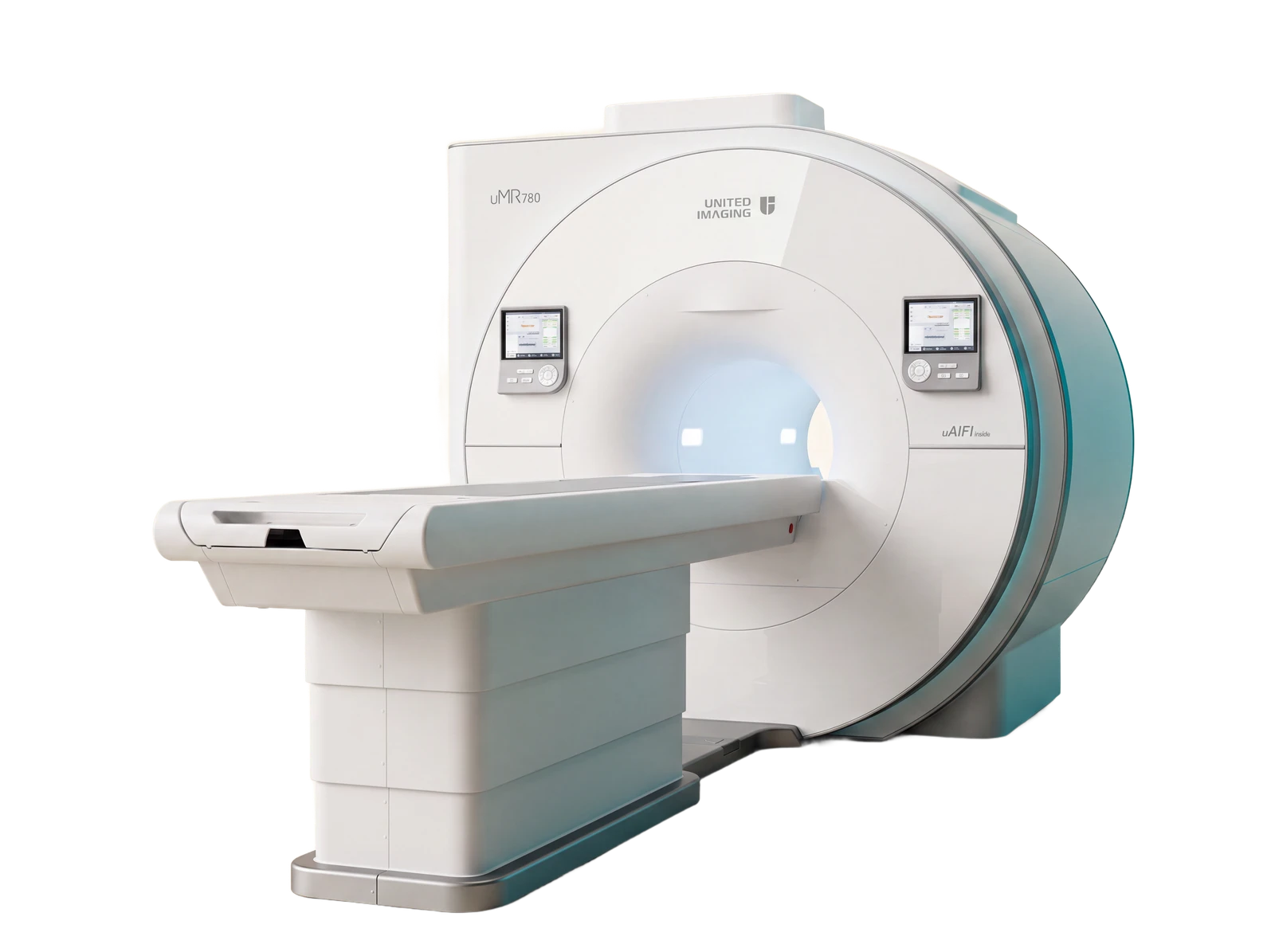
Detail that changes decisions, in a scan you can sit through.
What it is
A latest-generation 3 Tesla MRI platform - twice the field strength of a conventional 1.5T system - built on United Imaging's uCS acceleration engine. Higher signal means finer anatomical detail, and compressed-sensing acceleration of up to 16x shortens most studies without shortening the radiologist's read. The short-bore magnet keeps a full 50 cm imaging field while feeling less confining.
Clinical capabilities
- One 3D scan, every plane: MATRIX sequences are acquired once at 1 mm isotropic resolution and reconstructed in any plane - a T2 brain study in well under two minutes, the whole spine in just two acquisitions.
- Advanced neuro imaging for stroke, epilepsy and tumour work: diffusion tractography (DTI), susceptibility imaging, perfusion, functional BOLD and spectroscopy on the same magnet.
- Body MRI that outruns motion: whole-liver dynamic phases every 0.5 seconds across a single breath-hold, and MRCP that maps the bile and pancreatic ducts without endoscopy.
- Sub-millimetre joint imaging: PDW MATRIX at 0.6 mm isotropic separates ligament, cartilage and marrow detail - the sports-injury workhorse.
- MR angiography without ionising radiation, including non-contrast TOF studies and whole-brain vessel-wall imaging.
- Comfort engineering: motion-corrected sequences for restless patients and paediatric brain protocols measured in seconds, inside a wide, quieter bore.
16x
peak acceleration - whole-liver dynamic phases every 0.5 s in one breath-hold
0.6 mm
isotropic joint detail with PDW MATRIX sequences
2
acquisitions for a complete whole-spine study
1:42
minutes for a 1 mm isotropic 3D T2 brain sequence
Examinations it serves
- Brain & spine MRI
- Joint & sports-injury MRI
- Abdomen & pelvis MRI
- MR angiography
- Whole-body & small-parts studies
Platform capabilities as described by the manufacturer; specific features depend on the installed configuration - confirmed at the centre.
Seen on the uMR 780
Clinical images from the platform's application portfolio - the kind of studies this magnet was built for.
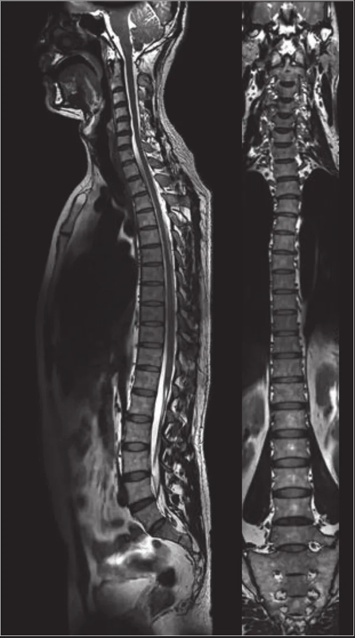
The whole spine in two acquisitions - cord and every disc level, without repositioning. 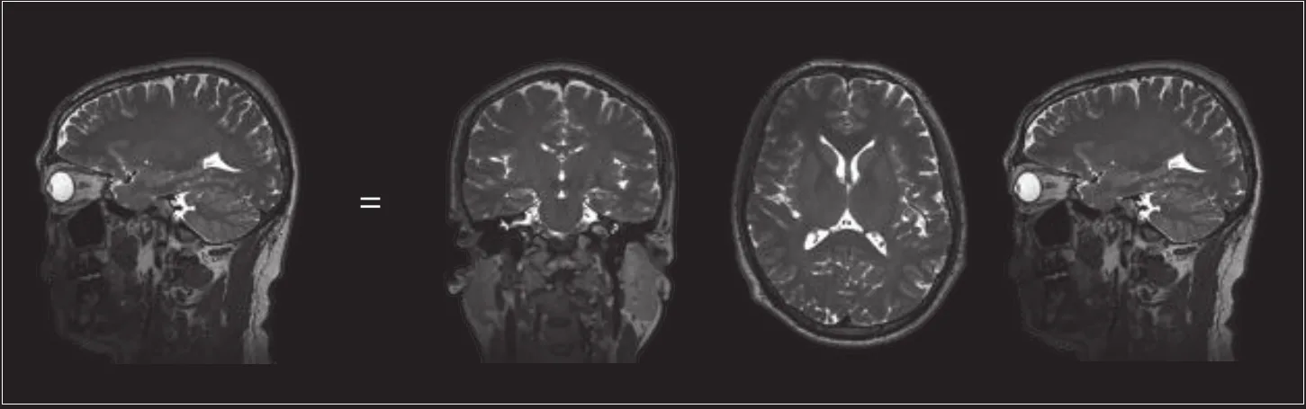
One isotropic T2W MATRIX acquisition (1 mm, 1:42 min) reconstructed in any plane - fewer repeats, more answers. 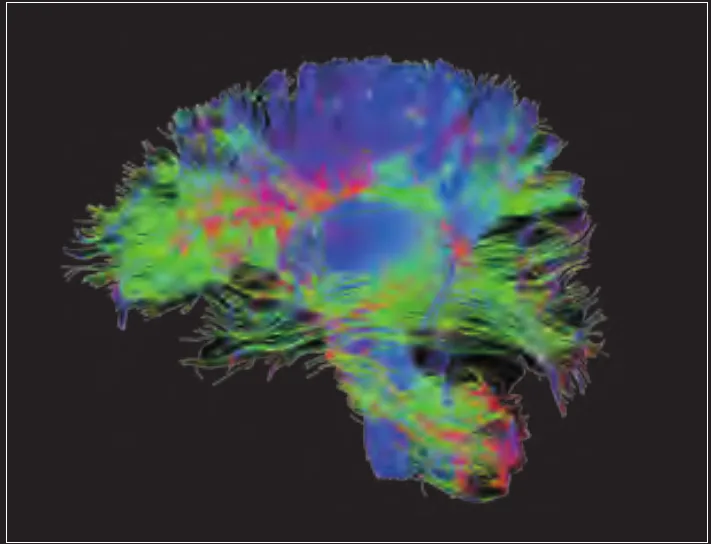
DTI tractography maps the brain's white-matter pathways - part of the advanced neuro toolkit alongside SWI, perfusion and spectroscopy. 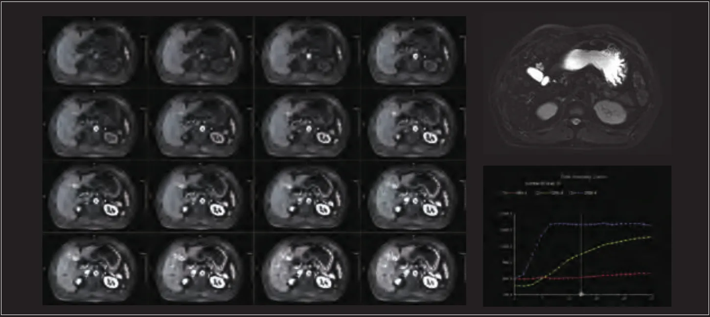
Whole-liver dynamic enhancement at uCS 16x - 32 phases at 0.5 s per phase in a single breath-hold, so no enhancement window is missed. 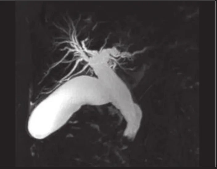
MRCP maps the bile and pancreatic ducts without endoscopy - T2 MATRIX at 0.9 x 0.9 x 1.2 mm. 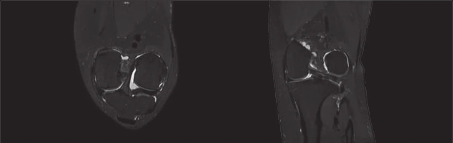
PDW MATRIX at 0.6 mm isotropic - the ligament, cartilage and marrow detail sports-injury medicine relies on.
Representative clinical images published by United Imaging for the uMR 780; acquisition details as stated in the manufacturer's portfolio. They illustrate platform capability, not patient studies performed at this centre.
02 · Siemens Healthineers
SOMATOM go.Now
CT
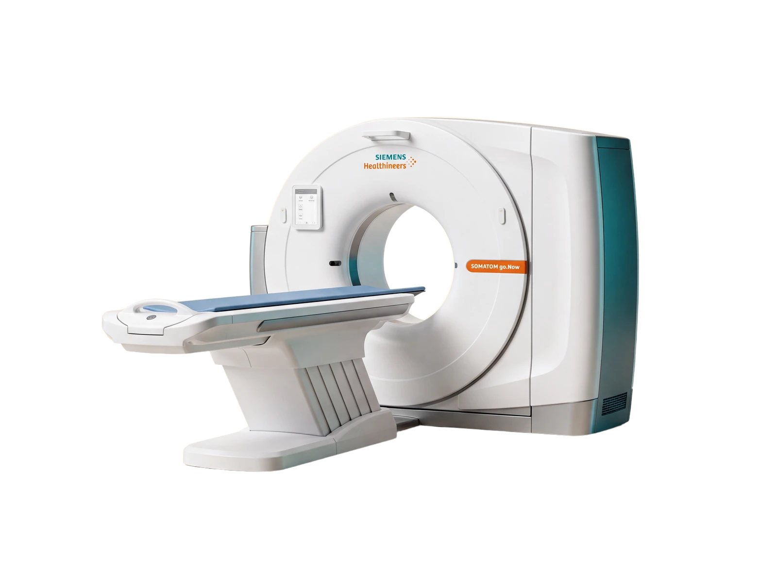
Answers in seconds, at the lowest dose the study allows.
What it is
A modern 32-slice CT built around dose discipline: Siemens' Stellar detector and Tin Filter technology keep radiation as low as reasonably achievable, while automated anatomy recognition plans and positions each scan consistently. The technologist controls the examination from a tablet, staying beside you.
Clinical capabilities
- Trauma, stroke and acute-abdomen imaging 24 x 7 - a full abdomen and pelvis covered in about 21 seconds, a contrast chest in about 14.
- Ultra-low-dose chest CT with the Tin Filter - lung studies at a fraction of conventional dose, with automated nodule measurement (diameter, volume, WHO area) for confident follow-up.
- Small-stone clarity in the abdomen: the Stellar detector resolves very small kidney and ureteric stones on 1 mm reconstructions.
- CT angiography from head to toe - automated anatomy recognition plans even whole-body runoff studies, 3D-reconstructed for surgical and clinical planning.
- Head CT tuned for the brain: strong grey-white matter differentiation, with X-CARE dose shielding that protects the eye lenses.
- iMAR metal artifact reduction for clear images around implants and prostheses - plus CT-guided biopsies and drainages with precise needle placement.
~14 s
for a contrast-enhanced chest acquisition in the published case series
1.34 mGy
CTDIvol of the ultra-low-dose Tin Filter lung case - dose discipline in practice
X-CARE
organ-based dose shielding that lowers absorbed dose to the eye lenses
iMAR
metal artifact reduction for clear imaging around implants
Examinations it serves
- Head & sinus CT
- Chest & lung CT
- Abdomen & pelvis CT
- Bone & trauma CT
- CT angiography
Platform capabilities as described by the manufacturer; specific features depend on the installed configuration - confirmed at the centre.
Seen on the SOMATOM go.Now
Cases from the platform's published clinical collection - what dose-disciplined CT looks like in practice.
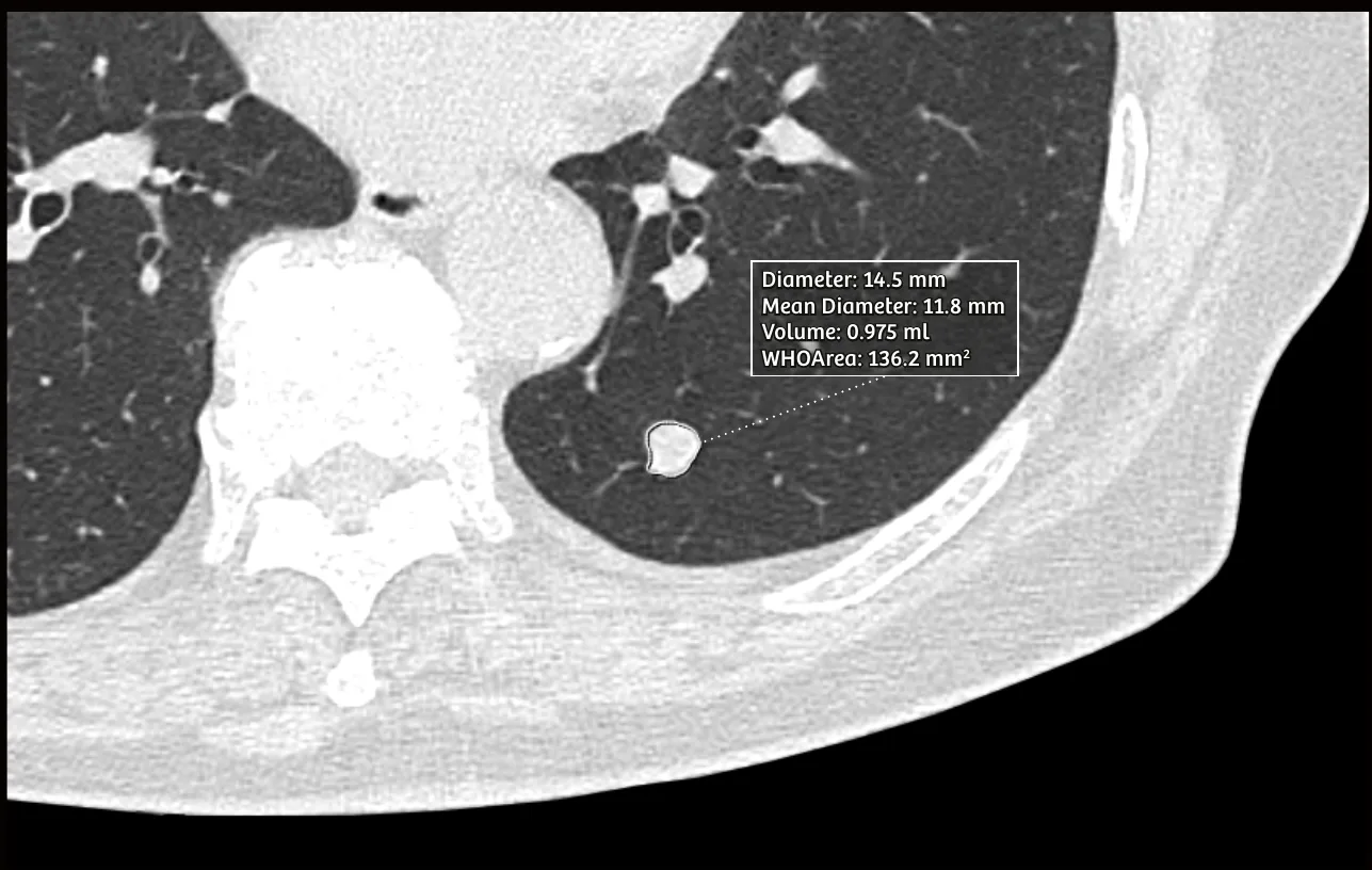
Automated nodule evaluation on a low-dose chest study - diameter, volume and WHO area measured in a click, so follow-up comparisons are exact. 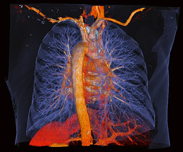
A 14-second contrast chest study rendered in 3D - airways, vessels and lung tissue from one acquisition at CTDIvol 5.29 mGy. 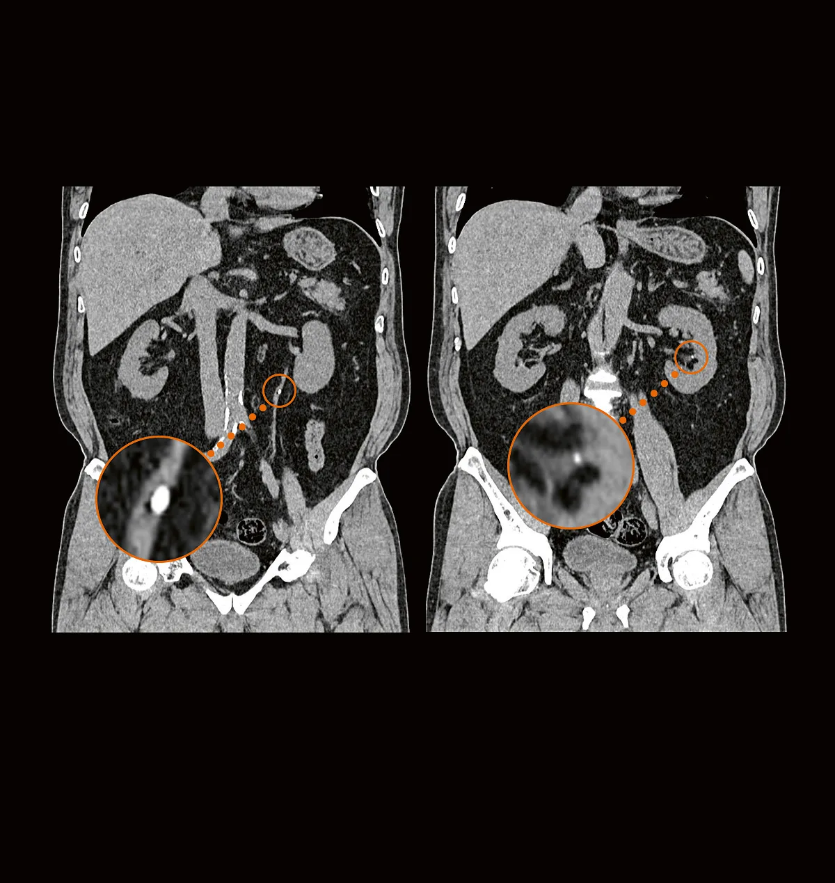
Very small urinary stones resolved on 1 mm coronal reconstructions - 458 mm of abdomen and pelvis covered in 21 seconds. 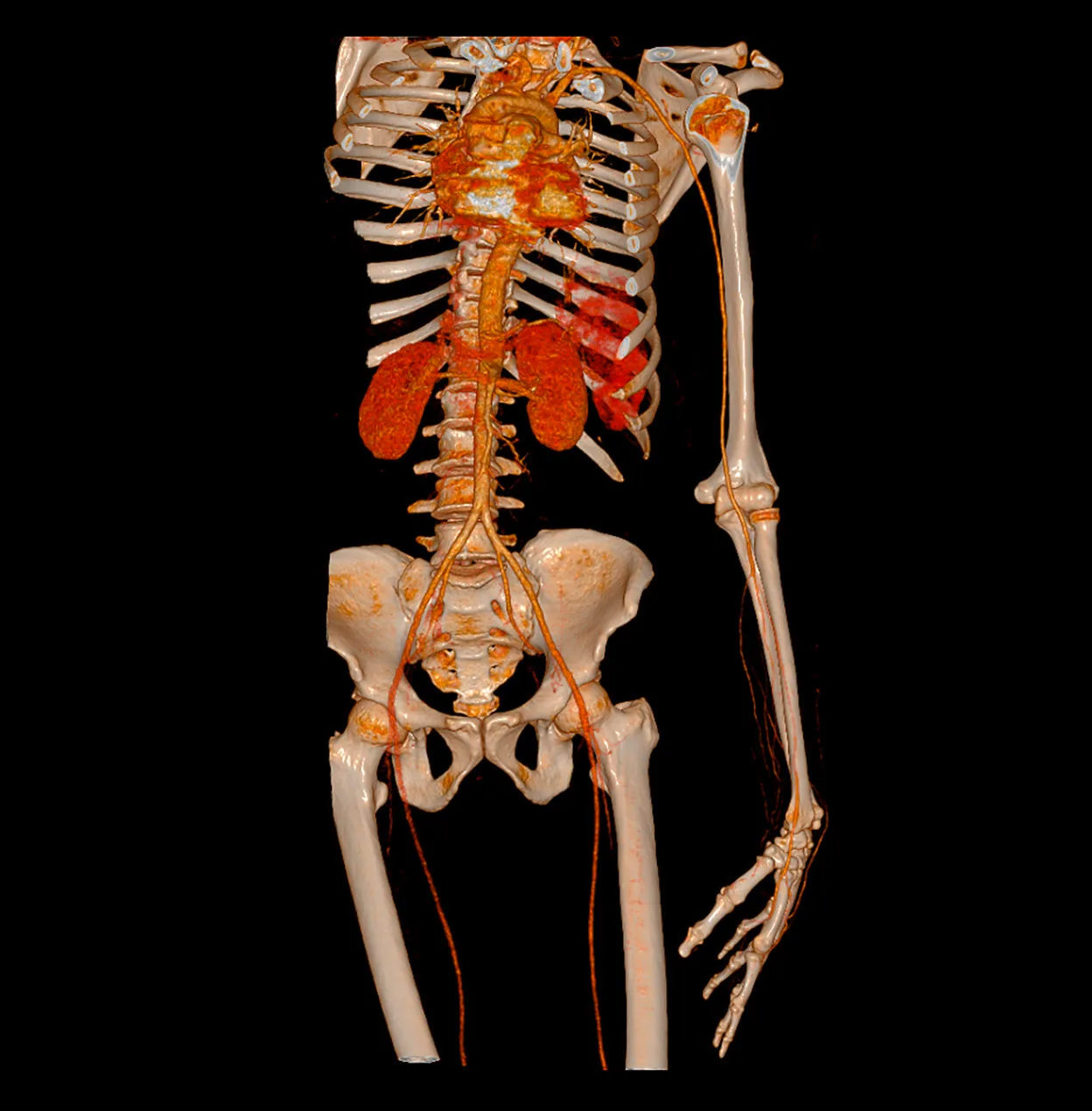
Whole-body CT angiography - 880 mm of vasculature in a 41-second scan, planned by automated anatomy recognition. 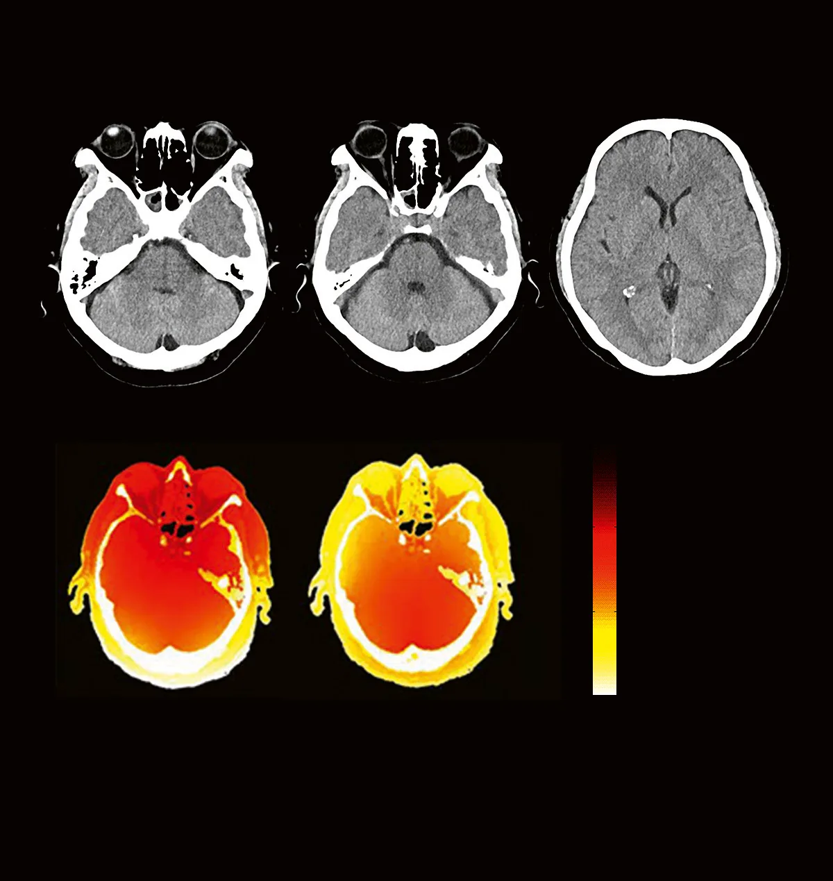
Grey-white matter clarity for the brain - and the dose maps behind it: X-CARE visibly lowers absorbed dose at the eye lenses. 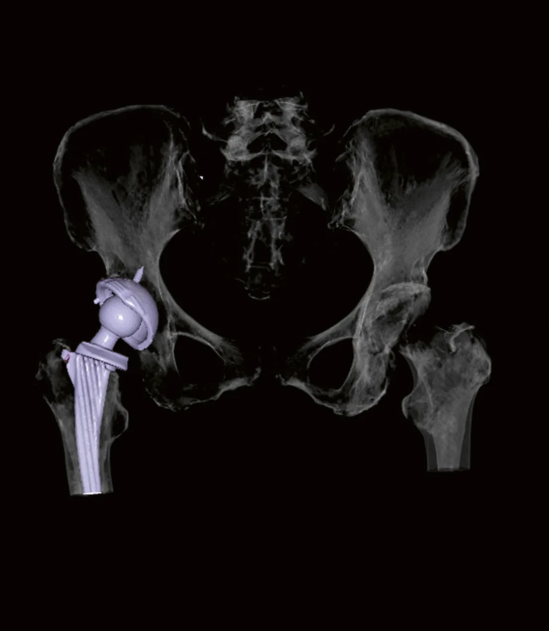
iMAR metal artifact reduction - a hip prosthesis rendered without metal bloom, so the bone around an implant stays readable.
Representative clinical cases published by Siemens Healthineers for the SOMATOM go.Now; scan parameters as stated in that collection. They illustrate platform capability, not patient studies performed at this centre.
03 · Philips
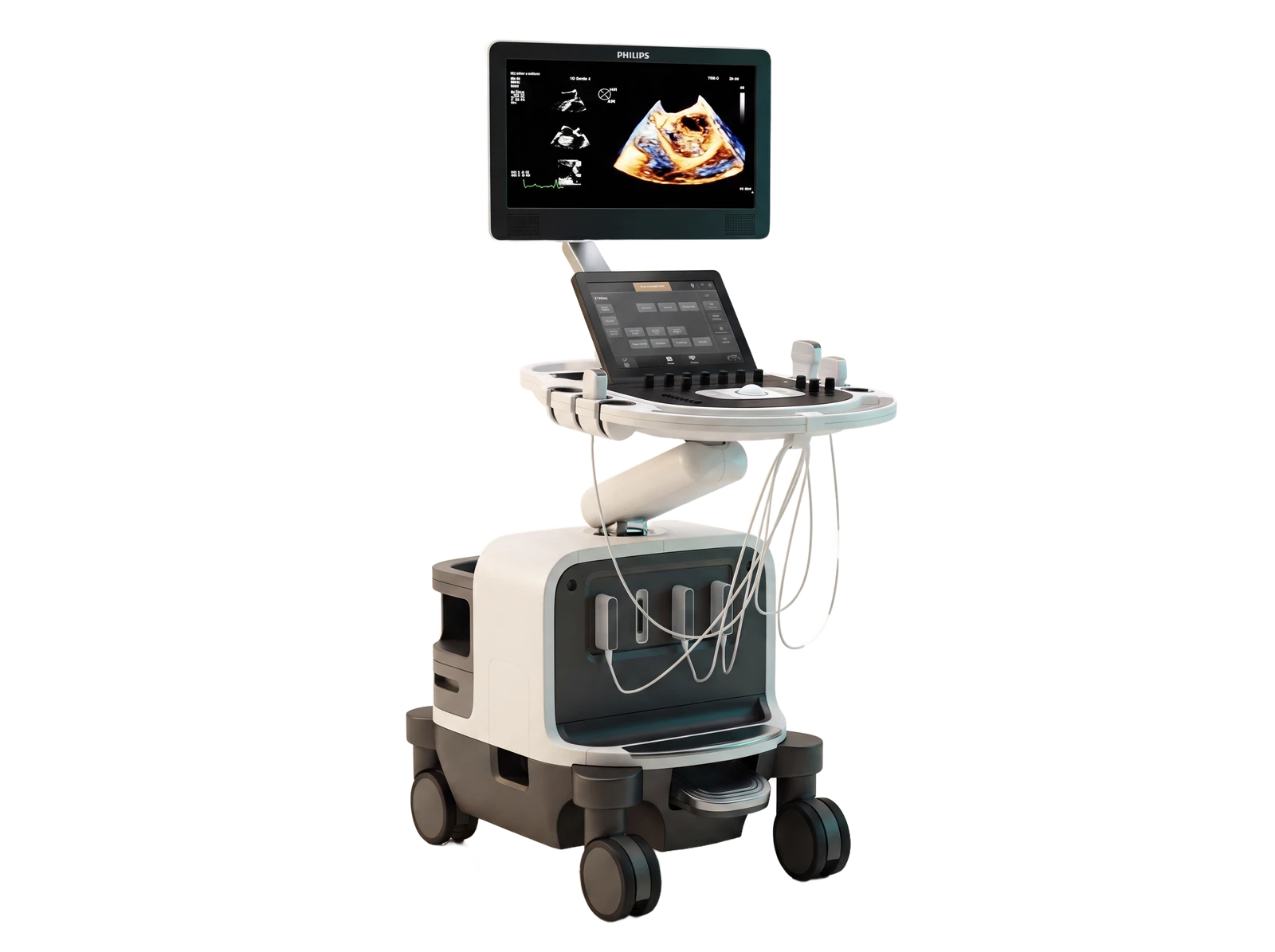
EPIQ 7
Premium Ultrasound
The most operator-dependent scan in radiology, done on the best platform available.
What it is
Philips' premium ultrasound platform. Ultrasound is the most operator-dependent examination in radiology - image quality depends on both the machine and the hands on the probe - so the centre pairs experienced sonologists with the EPIQ 7's PureWave single-crystal probes and high-sensitivity Doppler.
Clinical capabilities
- Shear wave elastography: liver stiffness and fatty-liver assessment without a biopsy.
- Microvascular Doppler that shows fine, slow blood flow - detail conventional Doppler can miss.
- Clear images even in difficult-to-scan patients - deeper tissue, post-surgical changes.
- 3D/4D imaging and fusion guidance, and real-time steering of image-guided procedures in the same visit.
Examinations it serves
- Abdominal & pelvic ultrasound
- Obstetric scans (PCPNDT-compliant)
- Thyroid, breast & small parts
- Vascular colour Doppler
- Ultrasound-guided procedures
Platform capabilities as described by the manufacturer; specific features depend on the installed configuration - confirmed at the centre.
United Imaging, Siemens Healthineers and Philips are trademarks of their respective owners.
Digital X-ray with videofluoroscopy, mammography and OPG complete the six modalities under this roof - platform details for these are published as the practice confirms them. See all imaging services
Book a scan
Want your study on these machines?
Request an appointment and our team will confirm timing and preparation with you personally.