Skip to content
Advanced
Imaging Point
The House of Imaging

Expertise

The scan is data.
The reading is the answer.

Advanced Imaging Point has always been a radiologist-led practice: imaging technology in service of careful, experienced interpretation.

01 · The radiologists

The people behind the reports.

A team of seven qualified radiologists, experienced across the major areas of diagnostic imaging.

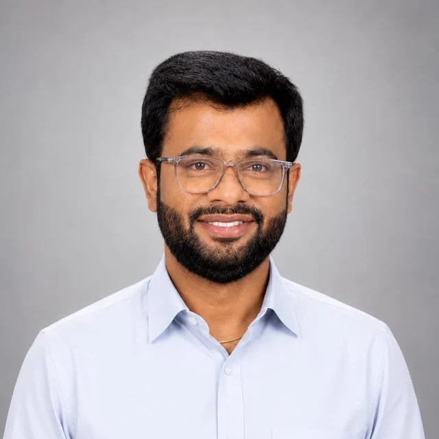
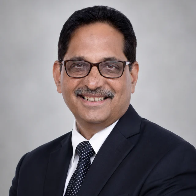
Portrait of Dr. R. R. Khandelwal

Chief Consultant Radiologist & Founder

Dr. R. R. Khandelwal

MBBS, MD, Fellowship in Color Doppler (Thomas Jefferson University, USA)

Ultrasonology and Doppler. Founded Advanced Imaging Point in 1989 and has guided its growth for more than three decades - a mentor to a generation of Nagpur radiologists.

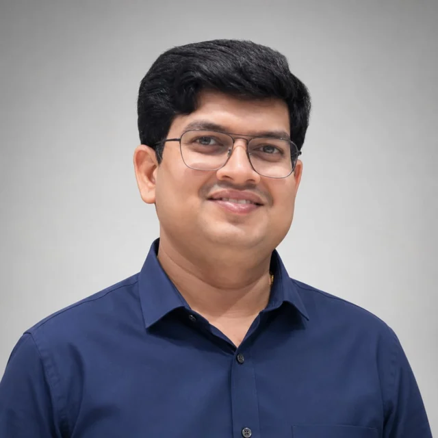
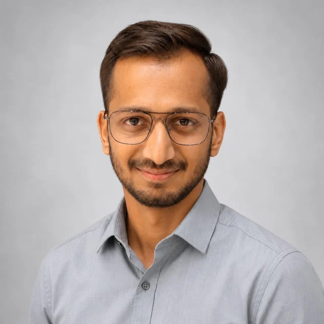
Portrait of Dr. Chetan Jaiswall

Consultant Radiologist

Dr. Chetan Jaiswall

MBBS, DMRD, DNB

Neuroradiology, body & abdominal imaging, and non-vascular interventions.

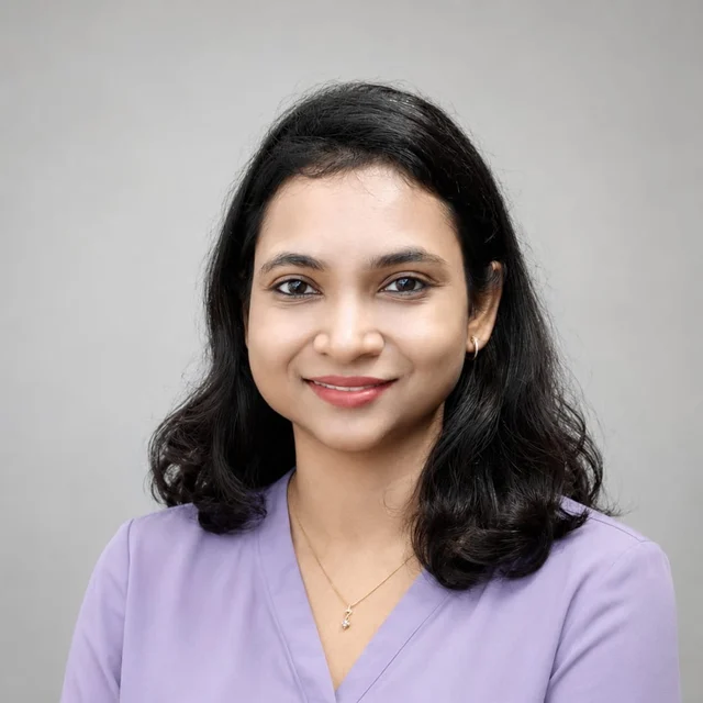
Portrait of Dr. Shefali Satardey

Consultant Radiologist

Dr. Shefali Satardey

MBBS, MD, DNB

Head & neck imaging, women's imaging and thoracic radiology.

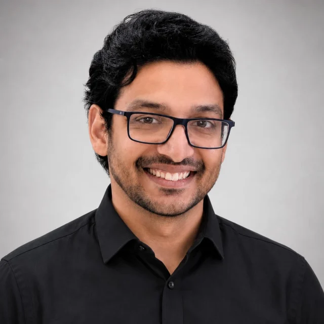
Portrait of Dr. Rachit Khandelwal

Consultant Radiologist

Dr. Rachit Khandelwal

MBBS, MD, EDiR, Fellowship in Musculoskeletal Imaging and Interventions (SMK, The Netherlands)

Musculoskeletal and neuro imaging, including image-guided MSK interventions.

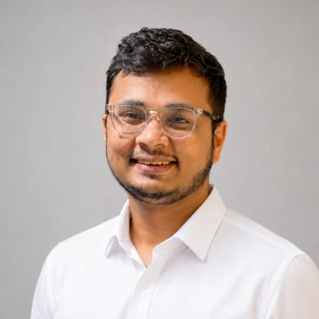
Portrait of Dr. Aditya Rakhunde

Consultant Radiologist

Dr. Aditya Rakhunde

MBBS, MD, Fellowship in Diagnostic Neuroradiology

Neuroradiology, fetal imaging and oncoimaging.

Portrait of Dr. Sandeep Chude

Consultant Radiologist

Dr. Sandeep Chude

MBBS, MD

Head & neck imaging, body imaging and non-vascular interventions.

Portrait of Dr. Sparsh Agrawal

Consultant Radiologist

Dr. Sparsh Agrawal

MBBS, MD

Musculoskeletal imaging, oncoimaging and body imaging.

Referring doctors are welcome to call and discuss any study with the reporting radiologist.

02 · Subspecialty areas

Eleven lenses, one practice.

Studies are read with the subspecialty lens they deserve - and complex cases can draw on more than one.

01 · Brain, spine, stroke & epilepsy

Neuroradiology

Brain and spine imaging with dedicated epilepsy and stroke protocols - including the sella, CP angle and temporal bone - read with the fine detail neurological questions demand.

02 · Sinuses, orbits, thyroid & neck

Head & Neck

Imaging of the sinuses, orbits, temporal bone, thyroid, salivary glands and neck spaces - for medical, dental and surgical planning.

03 · Joints, sports injury & bone

Musculoskeletal

Joints and sports injuries - cartilage and ligament imaging, small parts and bone tumours - imaged for injury, degeneration and unexplained pain.

04 · Liver, pancreas, renal & bowel

Body & Abdominal

Liver, pancreas, biliary tract, kidneys and bowel - including MR enterography and urography - supporting diagnosis, staging and follow-up.

05 · Chest & lungs

Thoracic

High-resolution chest CT for interstitial lung disease, nodule assessment and the full range of chest questions.

06 · Arteries & veins

Vascular

CT and MR angiography of the aorta, neck and limb vessels, with high-sensitivity colour Doppler for everyday vascular questions - as advised by your physician.

07 · Staging & follow-up

Oncologic

Staging and follow-up cross-sectional imaging with response assessment, reported for continuity so change over time is never missed.

08 · Breast, pelvic & obstetric

Women's Imaging

Breast imaging, pelvic MRI, and obstetric and gynaecological ultrasound - performed with privacy, and with strict adherence to the PCPNDT Act at every examination.

09 · Children's imaging

Paediatric

Age-appropriate protocols throughout, with dose-optimised CT - so children get the smallest exposure that answers the question.

10 · 24 x 7 acute imaging

Emergency & Trauma

CT and ultrasound available 24 x 7 for trauma, stroke and the acute abdomen - with round-the-clock reporting support.

11 · Image-guided procedures

Interventional (non-vascular)

FNAC, core biopsies, joint injections, fluid aspiration and pigtail drainage - performed in house under ultrasound or CT guidance.

Vascular interventional procedures are not performed in house - patients who need them are referred appropriately.
03 · The reporting process

How a scan becomes a report.

  1. 01

    Complete review

    Every study is read systematically, image by image - not skimmed for the obvious finding.

  2. 02

    Expert interpretation

    An experienced radiologist interprets the study in the context of your symptoms and your doctor's question.

  3. 03

    Correlation & comparison

    Previous imaging and clinical details are compared whenever they are available - change over time is often the diagnosis.

  4. 04

    Complex-case review

    Difficult or unusual studies may be discussed within the team, drawing on subspecialty experience where it helps.

  5. 05

    Clinical communication

    Significant or urgent findings are flagged for prompt communication with your referring doctor.

  6. 06

    A meaningful report

    The result is a clear, structured report written to be acted on - not decoded.

Every study receives a systematic read. Steps such as complex-case discussion and prior comparison apply where clinically relevant and where previous studies are available.

For referring clinicians

A phone call away, before and after the study.

Protocol queries, urgent requests, discussion of findings - call the centre and our team will connect you. Significant findings are flagged for prompt communication.

Book a scan

Ready when your doctor is.

Request an appointment and our team will confirm timing and preparation with you personally.